Ultrasound
Pelvic Ultrasound
A pelvic ultrasound is a noninvasive diagnostic exam that produces images that are used to assess organs and structures within the female pelvis. A pelvic ultrasound allows quick visualization of the female pelvic organs and structures including the uterus, cervix, vagina, fallopian tubes, and ovaries. This type of diagnostic testing may be used in patients who are experiencing infertility, abnormal uterine bleeding, pelvic pain, or postmenopausal bleeding, or those with a personal history of endometriosis, ovarian cysts, adenomyosis, or uterine fibroids. Pelvic ultrasound may be performed using one or both of the following methods:
- Transabdominal (through the abdomen) – a transducer is placed on the abdomen using conductive gel.
- Transvaginal (through the vagina) – a long, thin transducer is covered with the conducting gel and a plastic/latex sheath and is inserted into the vagina.
It is important to note if you are scheduled for a transabdominal ultrasound with our office and you are not pregnant, please be sure to drink 32 ounces of water one hour before your scheduled appointment time, without emptying your bladder. A full bladder helps by pushing bowel out of the pelvic region allowing transmission of ultrasound to target organs for better imaging.
Obstetric Ultrasound
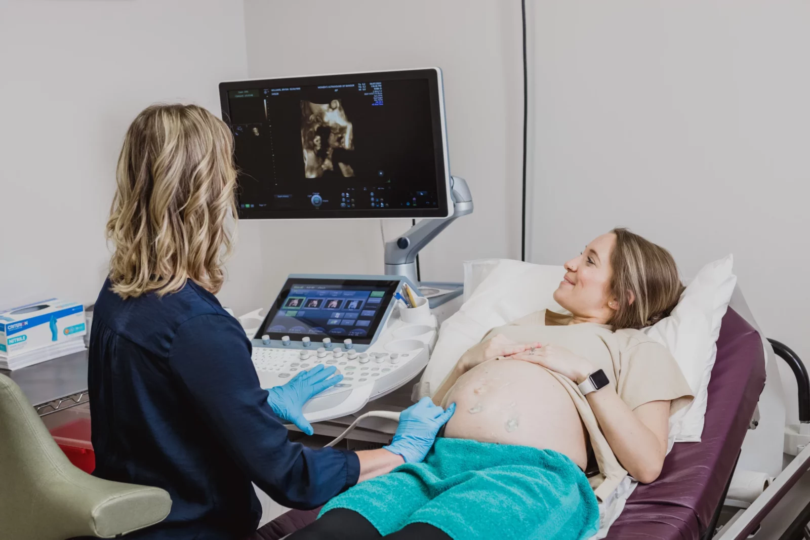
Routine obstetric ultrasound appointments:
8 weeks – transvaginal ultrasound to confirm intrauterine pregnancy, gestational age, and fetal heart
rate.
12-14 weeks – transabdominal ultrasound to measure nuchal translucency. Nuchal translucency (NT) is an ultrasound that measures the amount of fluid behind your baby’s neck. A small amount of fluid is normal, and measuring the amount of fluid can help calculate your baby’s chances of having a chromosomal or genetic variant. NT ultrasound is a screening test — it doesn’t diagnose a condition. It helps your healthcare provider determine if your baby is at risk and if further tests should be
recommended.
20 weeks – this is a 60-minute transabdominal ultrasound often referred to as the “anatomy scan”. Ultrasound at this gestational age confirms the gender, location of the placenta, and amniotic fluid volume. Fetal anatomy is assessed, and your sonographer will examine the following:
- presence of a four-chambered heart
- brain, neck, and spine
- kidneys and bladder
- arms and legs
- hands, fingers, feet, and toes
- lips, chin, nose, eyes, and face
- chest and lungs
- stomach and intestines
36-38 weeks – transabdominal ultrasound to reassess growth, weight, and presentation (breech/head-down) or a biophysical profile. A biophysical profile (BPP) helps observe the fetal heart rate, breathing, movement, muscle tone, and the amount of amniotic fluid.
It is important to note if you are scheduled for a prenatal ultrasound with our office and you are less than 33 weeks pregnant, you must arrive with a full bladder. This helps by pushing bowel out of the pelvic region allowing transmission of ultrasound to target the fetus for better imaging.